Clinical facilities
There are 2 treatment gantries and a dedicated treatment station for ocular tumours. These 3 treatment facilities used in clinical care can also be utilized for clinical studies or other R&D purposes. The imaging equipment consists of a CT scanner, a PET/CT scanner, and an MRI scanner. In addition, there is a research database with prospectively-collected patient information.
Treatment rooms
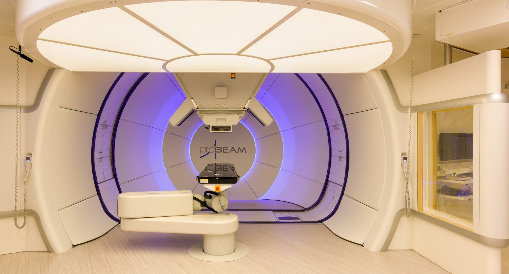
Treatment gantries
The 2 clinical proton therapy systems use pencil beam scanning technology in 360-degree gantries and are mounted with kV/cone-beam computer tomography (CBCT), for patient position verification. Furthermore, both treatment rooms are equipped with an in-room sliding Siemens SOMATOM Confidence CT scanner and a robotic bed to move the patient from imaging to treatment position, without altering the positioning.
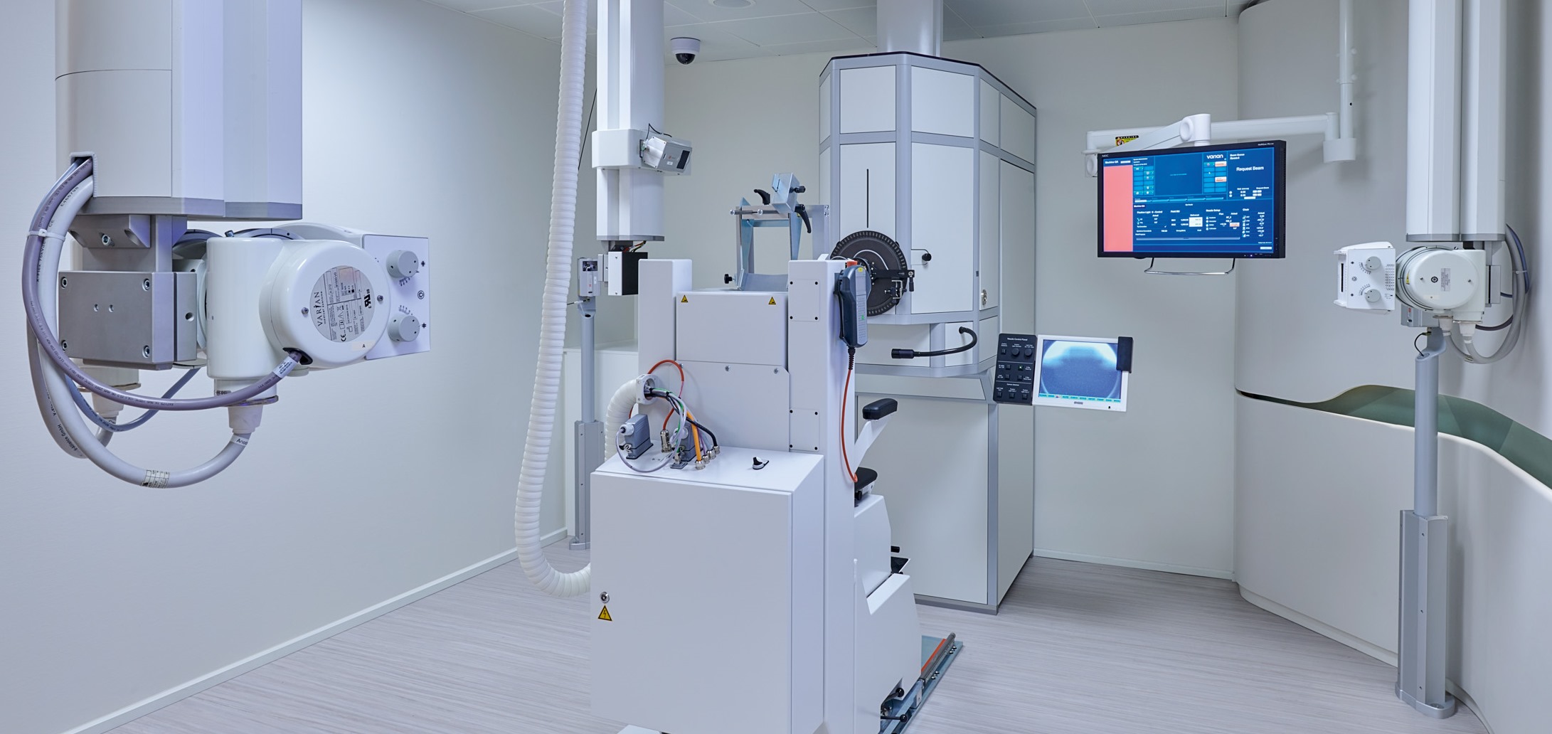
Ocular station
The ocular station consists of a fixed horizontal beamline and a single-scattering nozzle. It is fitted with orthogonal X-ray imaging equipment, utilizing digital flat-panel detectors, for patient position verification. A robotic chair allows positioning and immobilization of the patient with respect to the proton beam.
Imaging equipment
All imaging equipment is equipped with flat, hard-top couches and external lasers. Radiotherapy immobilization molds and indexing devices can be used.
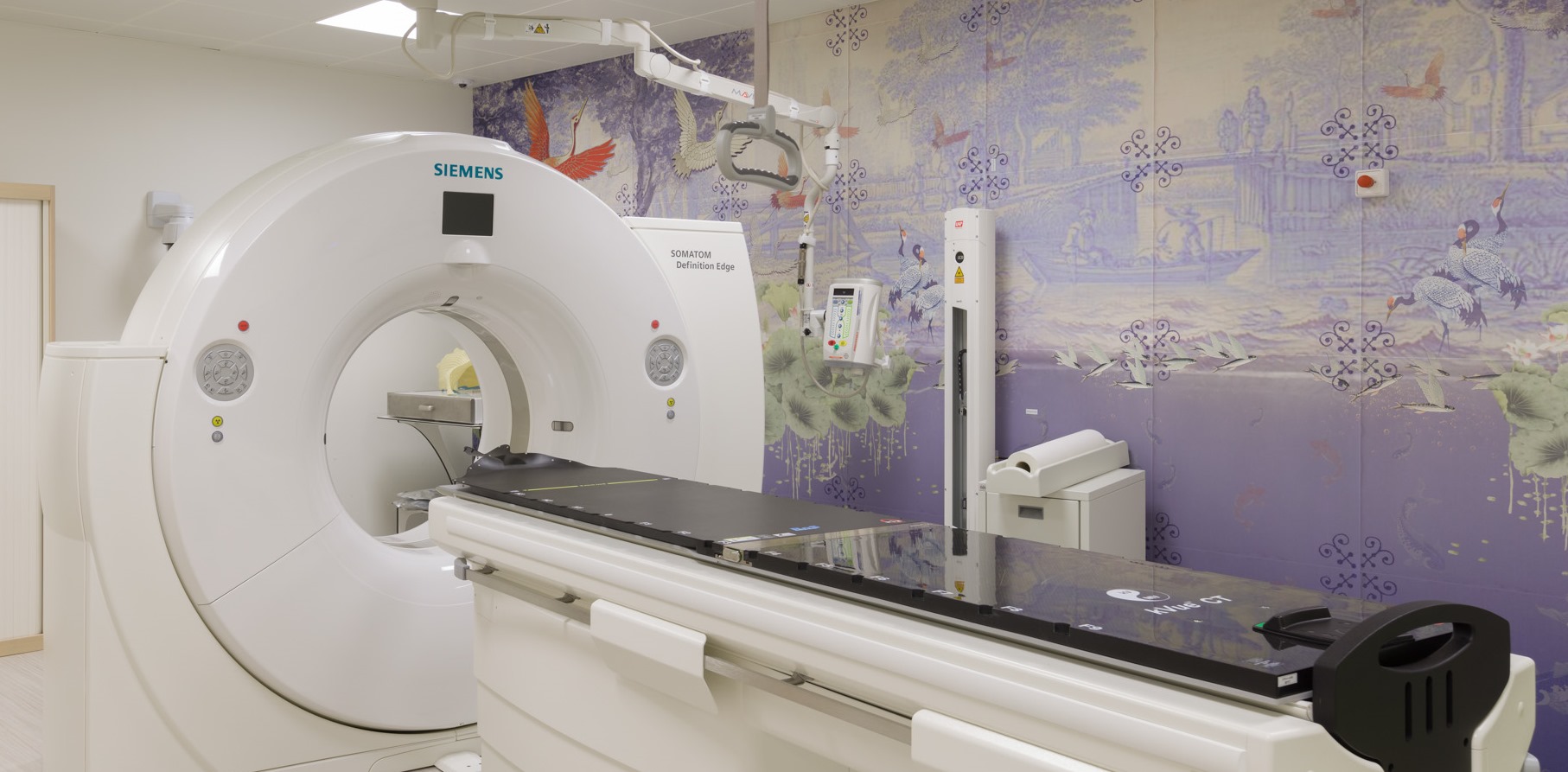
CT scanner
Siemens SOMATOM Definition Edge 128-slice dual-energy CT scanner, a single-source TwinBeam system with the option for simultaneous acquisition with 2 energy spectra for improved image quality and tissue characterization. This scanner is used for diagnostic imaging, treatment planning, treatment evaluation and treatment adaptation. It has 4D scanning options to detect motion and perform motion measurements.
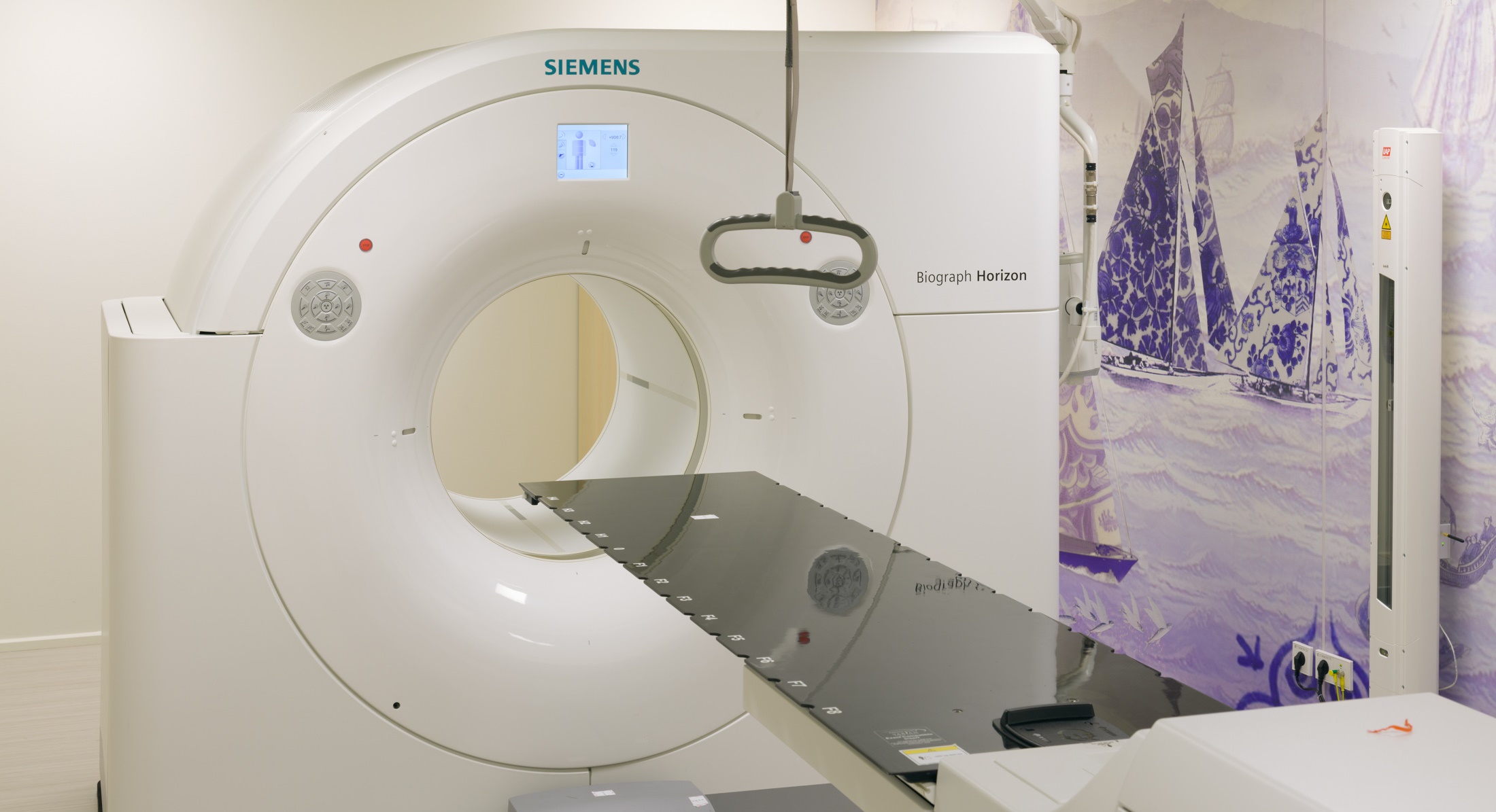
PET/CT scanner
Siemens Biograph Horizon PET/CT scanner with TrueV axial gantry extension, with the option for imaging in treatment position including patient set-up based on external lasers. The scanner is used for diagnostic imaging, treatment planning and treatment follow-up.
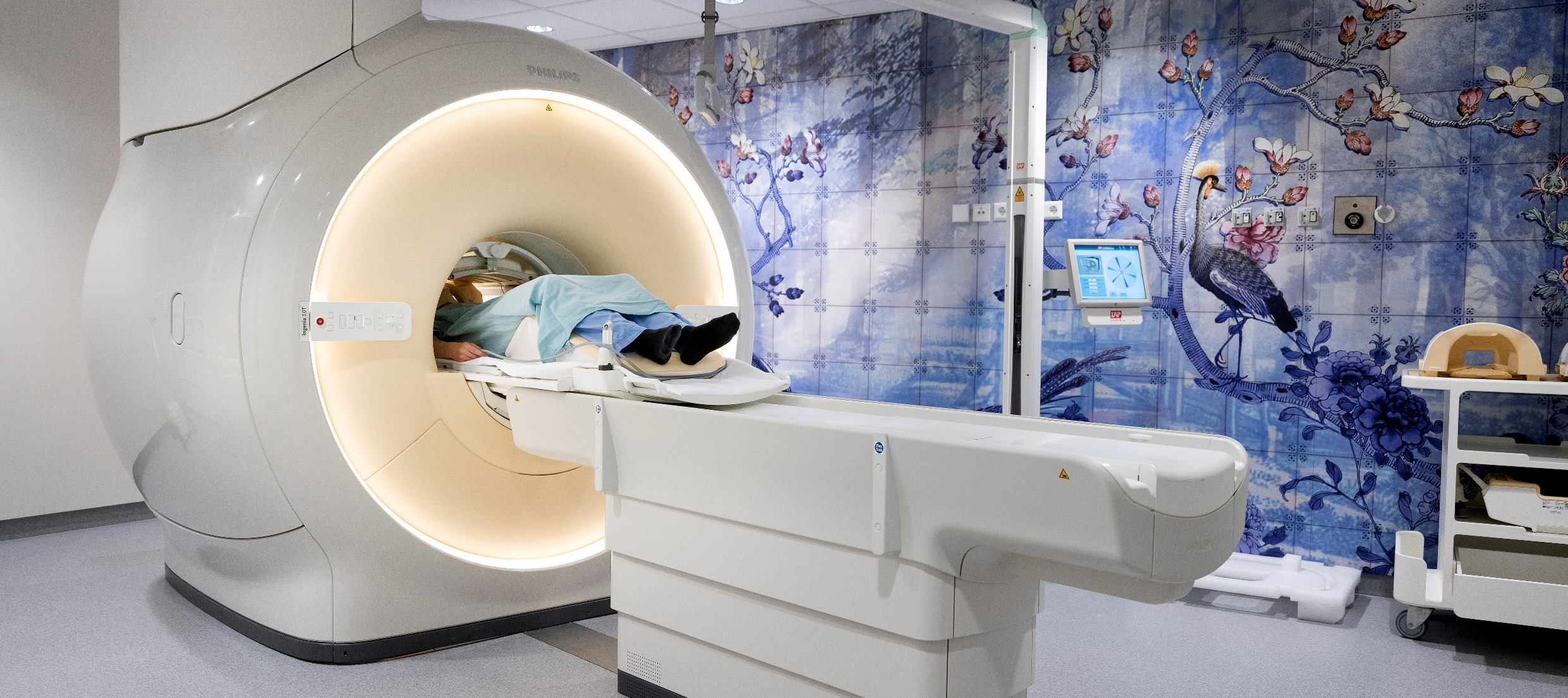
MRI scanner
Philips Ingenia MR-RT wide-bore 3T scanner dedicated for imaging in treatment position, including patient set-up based on external lasers. The scanner is used for diagnostic imaging, treatment planning and treatment follow-up.